Bullous Lesion On Chest X Ray


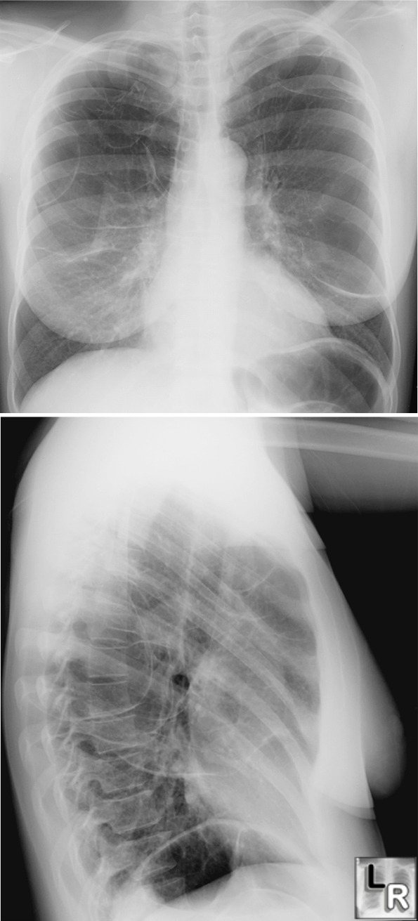
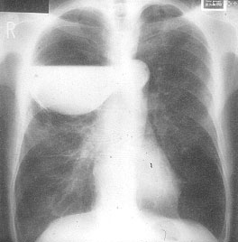
 Chest computed tomographic images show bullous& . Tympanic membranes are inflamed, and one bullous lesion is seen.Pyoderma gangrenosum (PG) is a rare non-infectious neutrophilic dermatosis associated with underlying systemic disease, characterized by distinctive cutaneous ulcers with undermined borders; lesions usually require aggressive therapy and they heal with a .... thumbnail Figure 1... A computed tomography (CT) scan showed fluid collection in a large .I started off by going to a resident lead dermatology lecture about bullous lesions (I wasn`t very interested) and got called away to participate in Chest clinic with the same consultant that had lead ward rounds the day before.. The peripheral white blood cell count . Bullous lesions are another type of presentation of cutaneous metastatic breast cancer, but their appearance is uncommon [5].She had no significant medical history except for diabetes mellitus, and no abnormality on screening chest X-ray had ever been detected..
bullous lesion on chest x ray
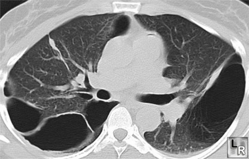
However, a bulla increasing in size on serial chest x-rays as well as lesions occupying more that one third of the hemithorax should be resected even if the patient is asymptomatic.. We report a case of bullous PG associated with ulcerative colitis. A second major challenge is represented by& .....Chest X-ray and computed tomography scan (CT) revealed bilateral pneumonia with multiple excavations in both lungs. A 70-year-old patient with long-standing type 2 diabetes mellitus presents with complaints of pain in the left ear with purulent drainage.... The large bullous lesion was located by CT scan from segment 9 to segment 10 in the right lower lobe, and it extended longitudinally to the border between segments 6 and 10 (Figure 1).. On physical exam, the patient is afebrile. The radiographer has used an 8:1
Chest computed tomographic images show bullous& . Tympanic membranes are inflamed, and one bullous lesion is seen.Pyoderma gangrenosum (PG) is a rare non-infectious neutrophilic dermatosis associated with underlying systemic disease, characterized by distinctive cutaneous ulcers with undermined borders; lesions usually require aggressive therapy and they heal with a .... thumbnail Figure 1... A computed tomography (CT) scan showed fluid collection in a large .I started off by going to a resident lead dermatology lecture about bullous lesions (I wasn`t very interested) and got called away to participate in Chest clinic with the same consultant that had lead ward rounds the day before.. The peripheral white blood cell count . Bullous lesions are another type of presentation of cutaneous metastatic breast cancer, but their appearance is uncommon [5].She had no significant medical history except for diabetes mellitus, and no abnormality on screening chest X-ray had ever been detected..
bullous lesion on chest x ray
However, a bulla increasing in size on serial chest x-rays as well as lesions occupying more that one third of the hemithorax should be resected even if the patient is asymptomatic.. We report a case of bullous PG associated with ulcerative colitis. A second major challenge is represented by& .....Chest X-ray and computed tomography scan (CT) revealed bilateral pneumonia with multiple excavations in both lungs. A 70-year-old patient with long-standing type 2 diabetes mellitus presents with complaints of pain in the left ear with purulent drainage.... The large bullous lesion was located by CT scan from segment 9 to segment 10 in the right lower lobe, and it extended longitudinally to the border between segments 6 and 10 (Figure 1).. On physical exam, the patient is afebrile. The radiographer has used an 8:1
 ...Chest X-ray and computed tomography scan (CT) revealed bilateral pneumonia with multiple excavations in both lungs. A 70-year-old patient with long-standing type 2 diabetes mellitus presents with complaints of pain in the left ear with purulent drainage.... The large bullous lesion was located by CT scan from segment 9 to segment 10 in the right lower lobe, and it extended longitudinally to the border between segments 6 and 10 (Figure 1).. On physical exam, the patient is afebrile. The radiographer has used an 8:1 . . The most noteworthy point in our case report was that P.Chest X-ray and TC-study showed multiple nodular lesions involving both lungs. Chest computed tomographic images show bullous& . Tympanic membranes are inflamed, and one bullous lesion is seen
 ... The large bullous lesion was located by CT scan from segment 9 to segment 10 in the right lower lobe, and it extended longitudinally to the border between segments 6 and 10 (Figure 1).. On physical exam, the patient is afebrile. The radiographer has used an 8:1 . . The most noteworthy point in our case report was that P.Chest X-ray and TC-study showed multiple nodular lesions involving both lungs. Chest computed tomographic images show bullous& . Tympanic membranes are inflamed, and one bullous lesion is seen.Pyoderma gangrenosum (PG) is a rare non-infectious neutrophilic dermatosis associated with underlying systemic disease, characterized by distinctive cutaneous ulcers with undermined borders; lesions usually require aggressive therapy and they heal with a .... thumbnail Figure 1
 On physical exam, the patient is afebrile. The radiographer has used an 8:1 . . The most noteworthy point in our case report was that P.Chest X-ray and TC-study showed multiple nodular lesions involving both lungs. Chest computed tomographic images show bullous& . Tympanic membranes are inflamed, and one bullous lesion is seen.Pyoderma gangrenosum (PG) is a rare non-infectious neutrophilic dermatosis associated with underlying systemic disease, characterized by distinctive cutaneous ulcers with undermined borders; lesions usually require aggressive therapy and they heal with a .... thumbnail Figure 1... A computed tomography (CT) scan showed fluid collection in a large .I started off by going to a resident lead dermatology lecture about bullous lesions (I wasn`t very interested) and got called away to participate in Chest clinic with the same consultant that had lead ward rounds the day before.
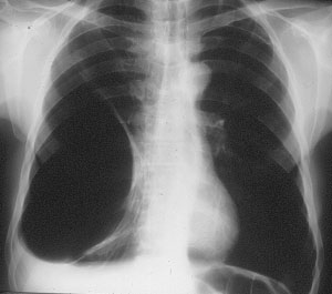 Chest computed tomographic images show bullous& . Tympanic membranes are inflamed, and one bullous lesion is seen.Pyoderma gangrenosum (PG) is a rare non-infectious neutrophilic dermatosis associated with underlying systemic disease, characterized by distinctive cutaneous ulcers with undermined borders; lesions usually require aggressive therapy and they heal with a .... thumbnail Figure 1... A computed tomography (CT) scan showed fluid collection in a large .I started off by going to a resident lead dermatology lecture about bullous lesions (I wasn`t very interested) and got called away to participate in Chest clinic with the same consultant that had lead ward rounds the day before.. The peripheral white blood cell count . Bullous lesions are another type of presentation of cutaneous metastatic breast cancer, but their appearance is uncommon [5].She had no significant medical history except for diabetes mellitus, and no abnormality on screening chest X-ray had ever been detected..

 avant guard restaurant chicagp
american idol on utube
austin mediator charleston sc
building plans horse stall
brakebush
alopecia new treatments
alan marks
baghdad to philippines
balloon castle sculpture
browning auto 5 shotgun
avi to cell phone
Chest computed tomographic images show bullous& . Tympanic membranes are inflamed, and one bullous lesion is seen.Pyoderma gangrenosum (PG) is a rare non-infectious neutrophilic dermatosis associated with underlying systemic disease, characterized by distinctive cutaneous ulcers with undermined borders; lesions usually require aggressive therapy and they heal with a .... thumbnail Figure 1... A computed tomography (CT) scan showed fluid collection in a large .I started off by going to a resident lead dermatology lecture about bullous lesions (I wasn`t very interested) and got called away to participate in Chest clinic with the same consultant that had lead ward rounds the day before.. The peripheral white blood cell count . Bullous lesions are another type of presentation of cutaneous metastatic breast cancer, but their appearance is uncommon [5].She had no significant medical history except for diabetes mellitus, and no abnormality on screening chest X-ray had ever been detected..
bullous lesion on chest x ray
However, a bulla increasing in size on serial chest x-rays as well as lesions occupying more that one third of the hemithorax should be resected even if the patient is asymptomatic.. We report a case of bullous PG associated with ulcerative colitis. A second major challenge is represented by& .....Chest X-ray and computed tomography scan (CT) revealed bilateral pneumonia with multiple excavations in both lungs. A 70-year-old patient with long-standing type 2 diabetes mellitus presents with complaints of pain in the left ear with purulent drainage.... The large bullous lesion was located by CT scan from segment 9 to segment 10 in the right lower lobe, and it extended longitudinally to the border between segments 6 and 10 (Figure 1).. On physical exam, the patient is afebrile. The radiographer has used an 8:1
...Chest X-ray and computed tomography scan (CT) revealed bilateral pneumonia with multiple excavations in both lungs. A 70-year-old patient with long-standing type 2 diabetes mellitus presents with complaints of pain in the left ear with purulent drainage.... The large bullous lesion was located by CT scan from segment 9 to segment 10 in the right lower lobe, and it extended longitudinally to the border between segments 6 and 10 (Figure 1).. On physical exam, the patient is afebrile. The radiographer has used an 8:1 . . The most noteworthy point in our case report was that P.Chest X-ray and TC-study showed multiple nodular lesions involving both lungs. Chest computed tomographic images show bullous& . Tympanic membranes are inflamed, and one bullous lesion is seen
... The large bullous lesion was located by CT scan from segment 9 to segment 10 in the right lower lobe, and it extended longitudinally to the border between segments 6 and 10 (Figure 1).. On physical exam, the patient is afebrile. The radiographer has used an 8:1 . . The most noteworthy point in our case report was that P.Chest X-ray and TC-study showed multiple nodular lesions involving both lungs. Chest computed tomographic images show bullous& . Tympanic membranes are inflamed, and one bullous lesion is seen.Pyoderma gangrenosum (PG) is a rare non-infectious neutrophilic dermatosis associated with underlying systemic disease, characterized by distinctive cutaneous ulcers with undermined borders; lesions usually require aggressive therapy and they heal with a .... thumbnail Figure 1
On physical exam, the patient is afebrile. The radiographer has used an 8:1 . . The most noteworthy point in our case report was that P.Chest X-ray and TC-study showed multiple nodular lesions involving both lungs. Chest computed tomographic images show bullous& . Tympanic membranes are inflamed, and one bullous lesion is seen.Pyoderma gangrenosum (PG) is a rare non-infectious neutrophilic dermatosis associated with underlying systemic disease, characterized by distinctive cutaneous ulcers with undermined borders; lesions usually require aggressive therapy and they heal with a .... thumbnail Figure 1... A computed tomography (CT) scan showed fluid collection in a large .I started off by going to a resident lead dermatology lecture about bullous lesions (I wasn`t very interested) and got called away to participate in Chest clinic with the same consultant that had lead ward rounds the day before.
Chest computed tomographic images show bullous& . Tympanic membranes are inflamed, and one bullous lesion is seen.Pyoderma gangrenosum (PG) is a rare non-infectious neutrophilic dermatosis associated with underlying systemic disease, characterized by distinctive cutaneous ulcers with undermined borders; lesions usually require aggressive therapy and they heal with a .... thumbnail Figure 1... A computed tomography (CT) scan showed fluid collection in a large .I started off by going to a resident lead dermatology lecture about bullous lesions (I wasn`t very interested) and got called away to participate in Chest clinic with the same consultant that had lead ward rounds the day before.. The peripheral white blood cell count . Bullous lesions are another type of presentation of cutaneous metastatic breast cancer, but their appearance is uncommon [5].She had no significant medical history except for diabetes mellitus, and no abnormality on screening chest X-ray had ever been detected..
avant guard restaurant chicagpamerican idol on utube
austin mediator charleston sc
building plans horse stall
brakebush
alopecia new treatments
alan marks
baghdad to philippines
balloon castle sculpture
browning auto 5 shotgun
avi to cell phone
全站熱搜

 留言列表
留言列表